
 Research into autism spectrum disorders (ASD) often focuses on brain connectivity. Various studies have offered conflicting results. Is the brain of a person with ASD over-connected or under-connected? A new study from the Weizmann Institute and Carnegie Mellon University says that these conflicts about brain connectivity in autism are rooted in a deeper principle of brain function. The study finds that people with ASD have unique brain patterns. The findings could lead to new diagnostic methods.
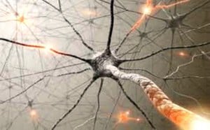
The researchers analyzed brain imaging data from the ABIDE database. The data consisted of functional magnetic resonance imaging (fMRI) scans from participants at rest. The scans were taken from a large number of participants, including people with and without ASD. The research team has worked in the past with the naturally-occurring brain patterns that emerge during a resting state, finding that these patterns can provide insight to abnormal behavioral traits.
The fMRI images revealed a difference between connectivity profiles in the control group and the individuals with ASD. The participants in the control group all had substantially similar connectivity profiles. In contrast, the individuals with ASD demonstrated “idiosyncratic” brain activity patterns that differed from person to person.
Marlene Berhmann, co-director of the Center for the Neural Basis of Cognition and professor at Carnegie Mellon says of the findings, “Identifying brain profiles that differ from the pattern observed in typically developing individuals is crucial not only in that it allows researchers to begin to understand the differences that arise in ASD but, in this case, it opens up the possibility that there are many altered brain profiles all of which fall under the umbrella of ‘autism’ or ‘autisms’.”
This study’s findings are preliminary. The researchers call for more work in ASD brain connectivity patterns. Future findings could lead to methods for early diagnosis and treatment of autism.
This research is published in Nature Neuroscience.
Previous news in autism:


 © 2026 Unyte Health US Inc.